Get Three-dimensional Images Of The Viscera With A Scanning Device

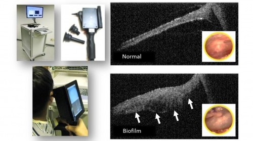
Engineers led by Stephen Boppart, a physician and biomedical engineer at from the University of Illinois Urbana-Champaign, have come up wit a hand-held scanning device that provides real-time three-dimensional images of the patients’ viscera. The team expects the scanner to be used in doctor’s offices or clinics in many ways: to assess hard-to-see-from-the-outside maladies such as ear infections, to examine diabetic patients, to monitor the health of their retinas. To image internal structures the scanner uses OCT system (optical coherence tomography), also known as “optical ultrasound,” cause it uses reflected light. The gadget also features a near-infrared light source, a video camera for obtaining images of surface features at the scan location, and a microelectromechanical systems (MEMS)-based scanner for directing the light. This project has lately received a US$5 million grant from the National Institutes of Health Bioengineering Research Partnership for elaborating the technology.
Via:gizmag.com
| Tweet |


